
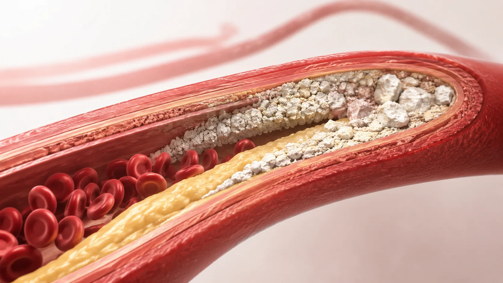
How Does Arterial Calcification Develop?
TL;DR: Arterial calcification develops gradually over years as calcium deposits accumulate in damaged or inflamed arterial walls. It is driven by vascular injury, inflammation, mineral imbalances, and aging — and while progression can be slowed through lifestyle and medical management, established calcification is very difficult to reverse.
What Is Arterial Calcification?
Arterial calcification is the buildup of calcium deposits within arterial walls, causing them to stiffen and lose elasticity. It is a well-recognised feature of cardiovascular aging and atherosclerosis, and it develops progressively over many years — often without symptoms until it is advanced. The process is not a single event but a slow, multifactorial cascade involving vascular injury, chronic inflammation, and dysregulated mineral metabolism.
There are two main subtypes. Intimal calcification occurs within atherosclerotic plaques and is strongly associated with inflammation and lipid accumulation. Medial calcification, in contrast, affects the middle layer of the arterial wall and is more closely linked to aging itself, diabetes, and chronic kidney disease. Both forms increase arterial stiffness and cardiovascular risk, though through somewhat different pathways.
The Biological Mechanisms Behind Calcification
How the Process Starts: Endothelial Injury and Inflammation
Calcification typically begins with injury to the endothelium — the thin inner lining of arteries. This damage can result from sustained hypertension, high circulating LDL cholesterol, smoking, oxidative stress, or hyperglycaemia. In response to injury, the body initiates an inflammatory reaction involving immune cells, cytokines, and reactive oxygen species. Over time, this chronic inflammatory environment sets the stage for calcium deposition.
Importantly, inflammation is not just a bystander. Evidence indicates that inflammatory signalling actively drives the cellular changes that lead to mineralisation within the vessel wall.
The Role of Vascular Smooth Muscle Cell Transformation
A key mechanism in arterial calcification involves vascular smooth muscle cells (VSMCs) undergoing a process of phenotypic transformation. Under conditions of oxidative stress and chronic inflammation, VSMCs can shift from their normal contractile role toward an osteoblast-like state — behaving more like bone-forming cells than vascular cells. As a result, they begin depositing calcium phosphate crystals in the arterial wall, in a process that closely resembles bone mineralisation.
This transformation is not fully understood, but it is strongly associated with metabolic syndrome, advanced age, and elevated phosphate levels. Targeting this pathway is an active area of cardiovascular research.
Calcium and Phosphate Dysregulation
Elevated serum phosphate, in particular, is a well-established driver of vascular calcification. This is especially relevant in chronic kidney disease, where impaired phosphate excretion leads to elevated circulating levels. When calcium and phosphate both rise, they tend to precipitate in soft tissues — including arterial walls — rather than remaining in bone where they belong.
Vitamin K2 plays a role in regulating this process by activating proteins that inhibit calcium deposition in soft tissue, though the clinical evidence for supplementation in preventing calcification remains limited in healthy populations.
Risk Factors and Contributing Conditions
Who Is Most at Risk?
Age is the single strongest independent predictor of arterial calcification. Beyond age, the major modifiable risk factors are hypertension, dyslipidaemia (particularly elevated LDL and ApoB), type 2 diabetes, smoking, and physical inactivity. Chronic kidney disease significantly accelerates the process due to its effects on mineral metabolism.
Individuals with metabolic syndrome — a cluster of elevated blood sugar, abnormal lipids, central obesity, and high blood pressure — carry a substantially higher risk. In these individuals, persistent low-grade inflammation and endothelial dysfunction create a sustained environment for calcification to develop.
For broader context on how individual organ systems age and interact, see our article on organ-specific aging.
How Chronic Conditions Accelerate Progression
Chronic diseases amplify the mechanisms described above. In diabetes, advanced glycation end-products (AGEs) damage the arterial wall and promote inflammation. In chronic kidney disease, dysregulated parathyroid hormone, elevated phosphate, and reduced active vitamin D all contribute to medial calcification. Hypertension increases mechanical wall stress, compounding endothelial injury over time.
These conditions do not simply add to normal aging — they can accelerate calcification by a decade or more, making early identification and management clinically important. The relationship between reduced kidney function and vascular calcification is explored further in our article on whether reduced eGFR increases mortality risk.
How Calcification Progresses Over Time
Arterial calcification develops silently over decades. Early-stage deposits are typically small and detectable only on imaging — often found incidentally during CT scans performed for other reasons. At this stage, there are usually no symptoms and no functional impairment.
As risk factors persist and inflammation continues, deposits grow and coalesce. The arterial wall becomes progressively stiffer, reducing its ability to absorb the mechanical stress of each heartbeat. This stiffening increases systolic blood pressure and pulse pressure, placing greater workload on the heart. In advanced cases, calcified plaques can destabilise or obstruct blood flow, increasing the risk of myocardial infarction or stroke.
The coronary artery calcium (CAC) score, measured by CT scan, is an established clinical tool for quantifying calcification burden and stratifying cardiovascular risk. It is one of the more reliable structural markers of subclinical cardiovascular disease and is used in clinical guidelines to inform treatment decisions — particularly around statin therapy in intermediate-risk individuals.
Can Calcification Be Reversed?
Once calcium deposits are established within arterial walls, reversal is extremely difficult. Current evidence does not support any widely available intervention that meaningfully reduces existing calcification. Most effective strategies focus on halting or slowing progression rather than reversing what has already accumulated.
Statins remain one of the most studied interventions. While they reduce cardiovascular events and slow atherosclerotic progression overall, they may actually increase CAC scores in some patients — likely by stabilising plaques rather than dissolving them. This does not mean statins worsen calcification in a harmful sense; rather, it illustrates the complexity of interpreting imaging findings in isolation.
Prevention and Management
Modifiable Lifestyle Factors
Lifestyle modification remains the most broadly applicable strategy for reducing calcification risk. Evidence supports the following approaches:
- Diet: Reducing ultra-processed food, managing dietary phosphate intake, and following an anti-inflammatory dietary pattern (such as a Mediterranean-style diet) can reduce vascular inflammation and support lipid control.
- Exercise: Regular aerobic exercise improves endothelial function, reduces blood pressure, lowers inflammatory markers, and supports insulin sensitivity — all of which reduce the conditions that drive calcification. Higher cardiorespiratory fitness is associated with lower CAC scores in population studies.
- Smoking cessation: Smoking is a direct cause of endothelial injury and accelerates atherosclerosis. Stopping smoking is one of the most impactful single interventions for vascular health.
- Blood pressure and blood sugar control: Managing both hypertension and hyperglycaemia reduces the mechanical and biochemical damage that initiates calcification.
Learn more in our complete guide to longevity.
Medical Management
For individuals with established risk factors or existing calcification, pharmacological management is typically indicated. Statins lower LDL cholesterol and reduce cardiovascular event risk regardless of calcification status. Blood pressure medications — particularly ACE inhibitors and ARBs — reduce arterial wall stress and have additional anti-inflammatory effects.
In patients with chronic kidney disease, phosphate binders and active vitamin D analogues are used to manage mineral dysregulation and reduce vascular calcification risk. These interventions are guided by established clinical protocols rather than speculative supplementation.
Overall, the most effective approach combines cardiovascular risk factor control with regular monitoring. For those at intermediate or higher risk, CAC scoring can provide meaningful prognostic information to guide treatment intensity.
References and Resources
Authoritative Sources on Arterial Calcification
-
American Heart Association – Atherosclerosis
heart.orgComprehensive overview of atherosclerosis development, the role of calcification, and associated risk factors and prevention strategies.
-
NIH – Pathogenesis of Vascular Calcification
nih.govDetailed review of the cellular and molecular mechanisms underlying arterial calcification, including VSMC transformation and mineral dysregulation.
-
European Society of Cardiology – Clinical Practice Guidelines
escardio.orgClinical guidelines covering cardiovascular risk management, including the role of imaging and pharmacological treatment in patients with calcification.
-
Circulation – AHA Journals
ahajournals.orgPeer-reviewed research on vascular calcification mechanisms, CAC scoring, and emerging therapeutic approaches.
-
National Kidney Foundation – Vascular Calcification
kidney.orgFocused resources on calcification in chronic kidney disease, including mineral management and its effects on vascular health.
-
Medscape – Vascular Calcification Overview
medscape.comClinical summaries, expert commentary, and updates on vascular calcification research and treatment options.
-
CDC – Cardiovascular Disease Facts
cdc.govEpidemiological data and public health guidance on cardiovascular disease, including arterial calcification as a contributing factor.
-
Nature Reviews Cardiology
nature.comIn-depth review articles covering the latest research on vascular calcification biology and investigational therapeutic strategies.
Frequently Asked Questions
How does arterial calcification develop?
Arterial calcification develops through a gradual process involving endothelial injury, chronic inflammation, and cellular transformation within the arterial wall. Damage to the artery lining — from factors such as hypertension, high cholesterol, or smoking — triggers an inflammatory cascade. Over time, vascular smooth muscle cells in the affected area begin to behave like bone-forming cells, depositing calcium phosphate crystals that progressively harden the arterial wall. This process typically unfolds over decades, often without noticeable symptoms in early stages.

Can arterial calcification be reversed once it develops?
Reversing established arterial calcification is not currently achievable through any widely available treatment. Most clinical interventions — including statins, blood pressure control, and lifestyle modification — aim to slow further progression and reduce cardiovascular event risk rather than dissolve existing deposits. Early detection and consistent risk factor management offer the best opportunity to limit long-term calcification burden.
What role do calcium and phosphate play in the development of calcification?
Elevated serum phosphate and disrupted calcium-phosphate balance are important drivers of vascular calcification, particularly in the context of chronic kidney disease. When these minerals are dysregulated, they tend to precipitate in arterial walls rather than remaining in bone. Managing phosphate levels through diet and, where necessary, medication is a clinically recognised strategy for reducing calcification risk in high-risk populations.
Which conditions accelerate arterial calcification?
Chronic kidney disease, type 2 diabetes, and hypertension are among the conditions most strongly associated with accelerated arterial calcification. Each promotes vascular injury, inflammation, or mineral dysregulation — the three core drivers of the process. Metabolic syndrome, which combines several of these factors, also significantly increases risk. Managing these conditions early and consistently is one of the most effective ways to reduce calcification progression.
How does aging influence arterial calcification?
Aging is one of the strongest independent risk factors for arterial calcification. With age, arteries accumulate cumulative wear from mechanical stress, oxidative damage, and low-grade inflammation — all of which promote calcium deposition. Some degree of age-related arterial stiffening is a normal physiological change. However, the rate and extent of calcification vary considerably between individuals and are meaningfully influenced by modifiable risk factors across the lifespan.
Conclusion
Arterial calcification is a well-characterised feature of vascular aging, driven by endothelial injury, chronic inflammation, cellular transformation, and mineral dysregulation. It develops slowly over many years and is strongly influenced by modifiable risk factors including hypertension, dyslipidaemia, diabetes, and smoking. While established calcification is very difficult to reverse, its progression can be meaningfully slowed through sustained lifestyle modification and appropriate medical management. Early detection — including coronary artery calcium scoring in appropriate individuals — supports more targeted risk stratification. Understanding how and why calcification develops is an important foundation for evidence-based cardiovascular prevention.
Find out more information about arterial calcification
Search for more resources and information: